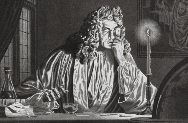
Perhaps one of the biggest ironies in biology is that microbes, which are the oldest self-replicating organisms on Earth, were among the last to be discovered and have largely been ignored. The history of their discovery is, like many in science, based on the invention of new technologies. In this case, it was a microscope created by a man named Anton van Leeuwenhoek. But before the Dutchman could make his serendipitous yet groundbreaking discovery in the late 17th century, lens-making technology had to turn several corners and see some other significant findings first.
In the 14th century, crude lenses were being fabricated in Europe for correcting vision. Toward the end of the 16th century, the Dutch began to use Venetian glass, the clearest and the highest quality glass available, to fashion relatively high-quality lenses.
Early in the 17th century, two Dutch lens-makers fashioned a telescope by pairing a concave and a convex lens in a tube. Although the instrument was not much more than a crude spyglass, having a magnification of about seven- or eightfold, it was a huge breakthrough in technology at the time.
In 1609, Galileo Galilei, using a telescope made in Italy from a Dutch lens-maker’s design, observed that the moons of Jupiter orbited that planet rather than the Earth. Although Galileo’s instrument had a magnification of only about twentyfold, it was sufficient to allow him to zoom in on what we already could see with our naked eyes: planets, stars and the moon. His observations threatened the prevailing Ptolemaic, or geocentric, understanding of the solar system and changed the way we think about our planet, ourselves and our special relationship to the universe.
Although stories of Galileo and his telescope abound, a somewhat lesser-known fact is that he also developed a microscope around 1619. It was simply an inadvertent outgrowth of the invention of the telescope, since it had been known for several years that by inverting a telescope with two lenses, one could magnify objects nearby. So, the optical design of the telescope was inverted and put into a new housing.
The microscope was smaller than its counterpart, and the two lenses were set in a barrel made of leather and wood. Regardless, Galileo did not seem to have much interest in what he saw with his inverted telescope. He appears to have made little attempt to understand, let alone interpret, the smallest objects he could observe. In fact, it was so irrelevant to him that he didn’t even give it a name. It wasn’t until 1625 that his peers decided to call it microscopio.
A Closer Look
The difference between the telescope and the microscope is not simply the configuration of lenses; it is also in human perception and the anticipation of what is seen. While the lack of perception may be partially due to hubris, I think most often it is the lack of looking for patterns in nature in places that are not normally accessible to our limited senses. We can see objects far away with our naked eyes. Comets, meteorites, planets, moons, stars and even exploding stars can be seen without a telescope, and hence, when they are brought closer for inspection with such an instrument as the telescope, these distant objects are not so mysterious, just somewhat so. However, our eyes cannot see something much less than the width of a hair (about a tenth of a millimeter) without the aid of a magnifying device. On the scale of microscopic structures, we are virtually blind. If we can’t even understand that there is a microbial world, why would we look for it?
The discovery of the microbial realm, like so many findings in science, was an accident that changed the world as profoundly as Galileo’s observations. It required a focusing of the mind as much as of an instrument. The breakthrough came in 1665, when the English Royal Society published the first popular science book, Micrographia (with the subtitle Or Some Physiological Descriptions of Minute Bodies Made by Magnifying Glasses With Observations and Inquiries Thereupon). It was written by Robert Hooke, then a 30-year-old hunchbacked, cantankerous, neurotic hypochondriac who was also a brilliant natural scientist, polymath and an original fellow of the society that published the book.
Micrographia captured many people’s imaginations. In it, along with dozens of beautiful engravings based on meticulous illustrations by the author, Hooke provided not only a clear description of the architecture of fleas, the seeds of thyme, the eyes of ants, the internal makings of sponges, microscopic fungi and the small building blocks of plants, but he also provided a detailed description of his own microscope.
Hooke’s observations were based on a relatively simple compound microscope that had two lenses. Instrument-makers at the time were familiar with telescopes and designed microscopes with two lenses, very similar to that of Galileo’s. But two-lensed microscopes had a big, unanticipated problem that telescopes did not. In such simple compound microscopes, the first lens created a halo of many colors that the second lens magnified. The result was that the more one magnified the object, the more distorted the image became.
The microscope Hooke used was well made by the standards of the time, but the optics were still poor. It suffered from the large optical aberration that lens-makers could not avoid. The best instrument, regardless of how lovingly the fabricator decorated it, could magnify an object only by about twentyfold before it became almost worthless. Even at such low magnification, the images were fuzzy, and sometimes a bit of imagination was required to reconstruct the structure of the object in view. Regardless, Hooke’s skillful illustrations were mind-boggling at the time, and the publication of Micrographia sparked interest in the construction of better lenses.
A Curious Find
It was around this time — in 1671, specifically — that Anton van Leeuwenhoek, a Dutch fabric merchant in Delft, developed a new but far less ornate microscope with smaller, simpler and, ironically, better optics that allowed much higher magnification without the distortion of the more complicated, expensive instruments. Rather than using two lenses, Leeuwenhoek pulled hot glass rods to form threads and then reheated the threads to form small glass spheres.
The glass spheres Leeuwenhoek used were about 1½ to 3 millimeters in diameter. There was a trade-off in the design of the lens: the smaller the lens, the higher the magnification, but also the smaller the field of view. He used the best Venetian glass and had to polish the lenses somehow. The exact technique he used was a secret he never revealed.
Leeuwenhoek constructed about 500 microscopes in his lifetime, and he had a variety on hand at any given moment to suit the purpose of what he was examining. The instruments themselves were relatively simple. A single spherical lens was mounted in a hole between a pair of silver plates. The sample was positioned on the back of the plates and was focused by a screw mechanism. The observer held the instrument up to his eye so that light from the sun or a candle could illuminate the object. The best instruments could magnify more than 200 times. This magnification was about equal to that of the microscope my father bought for me when I was 9 years old. Such instruments allow a human to see blood cells as well as animal sperm and single-celled organisms, including the “animalcules” that Leeuwenhoek observed. Indeed, it was the latter that would later be called microbes.
In October 1674, Leeuwenhoek fell ill, and he wrote (in Dutch), “Last winter while being very sickly and nearly unable to taste, I examined the appearance of my tongue, which was very furred, in a mirror and judged that my loss of taste was caused by the thick skin on the tongue.” He then went on to examine an ox’s tongue with his microscope and saw “very fine pointed projections” containing “very small globules.” He was describing taste buds. He then became curious as to how we sense taste and made infusions of various spices, including black pepper, in water.
In 1676, Leeuwenhoek found that a flask of pepper water that had been sitting on a shelf in his study for three weeks had become cloudy. In examining the cloudy water with one of his microscopes, Leeuwenhoek was surprised to find very small organisms swimming around. The organisms were only 1 to 2 micrometers in diameter — about one-hundredth the diameter of a human hair! He sketched the cells and wrote, “I saw a great multitude of living creatures in one drop of water, amounting to no less than 8,000 or 10,000, and they appear to my eye through the microscope as common as sand does to the naked eye.”
The discovery of animalcules was itself unforeseen. It was like seeing the moons of Jupiter but without a planet for the moons to orbit. It was a portent of untold numbers of invisible organisms and their presence right here on Earth. Leeuwenhoek had no idea what the organisms really were. He imagined they were literally extremely small animals, containing organs such as a stomach and a heart, just like the large animals we see with our naked eyes.
It is truly remarkable that the single-lens instruments made by Leeuwenhoek could allow him to see organisms so small, yet even with the best lenses of the day, he could not resolve their internal structures. However, Leeuwenhoek did something even more profound.
After the discovery of organisms in the pepper water, he examined scrapings from his own mouth. He was astonished to see, for the first time, the presence of animalcules on his teeth and gums. Here, Leeuwenhoek really stood out among the natural scientists; he revealed, for the first time, that we are not alone in our bodies. We are carriers of animalcules.
Indeed, animals like us harbor huge numbers of animalcules and help distribute them around the planet through our excretions and secretions. He also noted that when he drank hot coffee in the morning, the animalcules in his mouth died; it was the first observation that heat killed microbes. Leeuwenhoek went on to describe the various shapes and relative sizes of microbes he found in his own saliva and in other aqueous environments. His simple sketches would later become the basis for microbial taxonomy.
A Lasting Legacy
When Leeuwenhoek sent a 17-and-a-half-page letter to the Royal Society describing his discovery of animalcules for publication in the new, and first, scientific journal, Philosophical Transactions, it was met with such skepticism that even Hooke thought it was a delusion. Hooke sent an English vicar and some other reputable observers vetted by the Royal Society to Delft to verify the reports. The observers were as amazed as Hooke and his colleagues in London had been.
In 1677, Leeuwenhoek’s now-verified observations were published by the Royal Society (in English, after being translated from Dutch with help from Hooke, who learned Dutch so that he could read Leeuwenhoek’s papers). Leeuwenhoek was elected a foreign fellow of the society in 1680, but he never visited London. His descriptions and enumeration of microbes seemed to support the idea of spontaneous generation of life (in pepper infusions no less!), the idea that organisms could be formed from dead or nonbiological sources without any obvious parental lineage. For example, it was commonly accepted that maggots could form in dead meat and that wasps could come from buried elk horns. Spontaneous generation was widely believed by most people at the time. Leeuwenhoek rejected the basic notion, but he could not disprove it.
Even though Leeuwenhoek could not disprove the notion of spontaneous generation of life, his findings showed that he was a creative genius. He had no formal higher education and no affiliation with any university. He did not know Latin or Greek, the two languages of formally educated people at the time; he wrote only in Dutch. He built microscopes as a pastime and gave many of them away; he never sold any. He bequeathed 26 of his instruments to the Royal Society, all of which subsequently were “borrowed” by members of that esteemed group of scientists; all the originals have since disappeared. The rest of his collection was sold for the weight of the silver or other metals in the bodies of the instruments. Over his 90-year lifetime, he sired five children. Only one, Maria, lived beyond childhood, and his scientific legacy almost died with his own death in 1723.
Although Leeuwenhoek is often viewed as the father of microbiology, Hooke was the collaborative agent who led him to fame. Both remarkable men were critical catalysts for the impending discovery of the invisible world. On a personal level, both were extremely generous toward each other to the end of their lives.
Neither Hooke nor Leeuwenhoek had students, and although Micrographia was a big seller in 1665 and for some years afterward, Leeuwenhoek never wrote a book, and his papers were not widely read. Neither Leeuwenhoek nor Hooke had a biological successor, and unlike Galileo, neither had immediate intellectual successors.
Interest in pepper water faded. The role of microbes in biological functions was virtually ignored, and it would be almost 200 years before these organisms would garner further serious attention.
Amazingly, while the fundamental discoveries in science in the 17th century — gravity, light waves, planetary rotation around stars and the incredible abstraction of science in mathematics — spurred huge explosions of discoveries in physics and chemistry, fundamental discoveries in biology largely lagged behind and were important only as they related to human health. And so the microbial world was delegated to an invisible world in the 18th century — as natural philosophers turned to questions about the evolution of plants and animals, and the geologic structures that contained fossil remains of extinct organisms.
Excerpted from Life's Engines: How Microbes Made Earth Habitable by Paul G. Falkowski © 2015 by Princeton University Press. Reprinted by permission.

