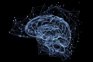
Mapping the structure of the human brain is a daunting task: Billions of cells make vast numbers of connections, and tracing these tangled fibers is so labor-intensive that analyzing just one square millimeter of tissue takes years.
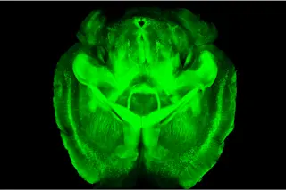
Into this murky mess, Stanford University neurobiologist Karl Deisseroth has brought CLARITY, a technique that turns brain tissue transparent while maintaining its structure. The method, described in Nature in April, makes it possible to inspect the 3-D architecture of an intact mouse brain in microscopic detail.
Traditionally, scientists explore neuroanatomy in animals by injecting dyes or stains that illuminate specific nerve cells and connections. They then kill the animal and slice its brain tissue thin enough so that light can shine through it under a microscope, revealing the structure within each slice.
But reconstructing 3-D architecture from a stack of slides is imprecise and slow. And neuroanatomists have been unable to look at ...


