Mysteries of the Heart

No one knows why a puzzling growth sometimes chokes off the blood vessels that embrace the heart, or who is most susceptible to it. But a seemingly harmless--and nearly ubiquitous--virus may provide a valuable clue.

By Steven Dickman
Jul 1, 1997 5:00 AMNov 12, 2019 6:26 AM

Newsletter

Sign up for our email newsletter for the latest science news
 

Of all the thousands of diseases that keep hypochondriacs awake at night, cancer of the heart is not one of them. Unlike bones, skin, brain, liver, blood, kidneys, and bowels, our heart muscle and the coronary blood vessels that embrace it rarely turn cancerous.

Stephen Epstein knows why cancer of the heart is so rare. As chief of the cardiology branch at the National Heart, Lung, and Blood Institute, Epstein has made it his business to know the heart inside out. Cancer occurs when cells grow out of control, he says. But in the heart and the vessels, cells are not growing very much. The cells divide only maybe once a year. No division, no cancer.

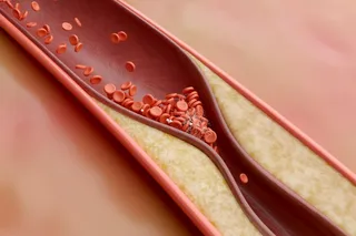
But there is one malady in which cells in the heart’s blood vessels do divide excessively, and its cause is a medical mystery. Every year, some 1 million Americans develop atherosclerosis, a partial or total blockage of the arteries. The coronary arteries that wrap around the heart and supply it with blood are especially vulnerable. Coronary atherosclerosis is not the mystery, however; its risk factors are relatively well documented, if not fully understood--junk food and Americans’ sedentary lifestyle on the one hand, genetics and metabolism on the other. The mystery is the gumming up of coronary arteries that all too often follows treatment for coronary atherosclerosis. And it is a mystery that Epstein would love to solve.

When coronary arteries narrow--and in doing so foreshadow a heart attack--doctors typically turn to angioplasty. A cardiologist threads a slender tube tipped with a deflated balloon into the blocked artery, inflates the balloon, and--voilà--blockages are squished against the sides of the artery and blood begins to flow again. No anesthesia is necessary, and the patient can often go home the same day. So far, so good. When angioplasty was first introduced, in the mid-1970s, it seemed like a miracle cure. The procedure was simple and far less invasive than grafting blood vessels to replace the diseased coronary arteries.

Inexplicably, though, angioplasty fails almost as often as it succeeds. Sometimes it takes weeks, sometimes months, but in 25 to 50 percent of the 400,000 angioplasties U.S. cardiologists perform each year, the arteries reclog. For reasons unknown, the smooth muscle cells that line the artery walls often grow excessively following angioplasty, choking off blood flow once more. The phenomenon, called restenosis, resembles the gradual narrowing of arteries but happens much, much faster. And as is not the case with atherosclerosis and its known accompanying risk factors, doctors have no way of predicting which patients’ arteries will gum up.

By the early 1990s, restenosis was more than an occasional medical condition, despite the emergence of a host of mechanical and chemical alternatives to angioplasty. One treatment Roto-Rootered away blockages with pulsating miniature switchblades. Another, more successful strategy jammed open arteries with stents, tiny coiled devices that look like the springs from ballpoint pens. Toying with angioplasty--altering the size and speed of the balloon expansion--didn’t solve the problem either. And 50 different drug treatments have failed to hold off the tightening noose of restenosis.

Why, Epstein wondered, do some patients’ arteries reclog and not those of others? Why do any of them reclog at all? And what does this condition tell us about the nature of coronary artery disease in the first place?

A few years ago Epstein concluded that the answers would only be found inside cells. And to look inside cells would require a detour outside the traditional boundaries of cardiology. Cardiac surgery made great inroads in the 1960s, he says. Then in the 1970s, angioplasty came along. But by the end of the eighties, it was clear that the major breakthroughs in surgery were no longer coming. So in 1990, Epstein decided it was time to redirect his career.

My tennis partner, Ira Pastan, was a world-class molecular biologist in cancer, Epstein recounts. I was telling him that I was looking for something new, and right there on the court, he offered me a bench in his laboratory for a sabbatical year.

Molecular biologists probe the machinery of cells themselves, a little like auto mechanics who look at carburetors and cylinders to understand engines. Cancer researchers had long perceived the usefulness of studying the cells’ components, not least because cancer often seemed to arise as the result of faulty communication among these moving parts. But because cardiologists had been so successful on the macro level, repairing valves and replacing vessels, they had for the most part stopped short of studying the molecules that make up hearts and arteries.

Switching fields was not easy, says Epstein. Here I was, 55 years old, learning molecular biology. At every lab meeting, I felt as if I was in the middle of Moscow. I didn’t understand what anyone was saying. Still, Epstein knew he’d made the right choice. In Pastan’s laboratory he could study the innards of the muscle cells that line artery walls. The excessive growth of such cells following angioplasty reminded Epstein--as it had other researchers before him--of cancer. Epstein and his co-worker, cell biologist Edith Speir, began hunting for the molecules that might be causing the growth.

By 1992, back in Epstein’s laboratory, they had selected their target: a protein called p53. This protein plays two important roles in regulating cell growth. First, it acts as a brake on cellular multiplication: when a cell’s genetic material is damaged or threatened, the cell churns out p53 to prevent the production of offspring until the damage can be repaired. And second, when a cell senses that its innards are somehow beyond repair, p53 flips a suicide switch that initiates the cell’s destruction.

A common hallmark of a cancerous cell, researchers had learned, is high levels of defective p53. In a normal cell p53 interacts with DNA-- somehow blocking inappropriate cell division--and is then degraded. But in a cancer cell with defective p53, the protein can’t do its job and it begins to accumulate. So elevated p53 levels became synonymous with cancer.

Epstein and Speir’s first task was to look for p53 in tissue samples from patients whose angioplasties had led to restenosis. Sure enough, there were large amounts of p53 in many of the samples. The next finding, however, left them bewildered: the p53 they’d isolated was not mutated but normal. Prevailing dogma held that just the opposite should be true.

If the protein was normal, why wasn’t it working to rein in cell growth? When we searched through the oncology literature, says Epstein, we noticed that very often, in the cancers in which p53 protein was overabundant but unmutated, there was evidence that the cells had become cancerous because viruses had interacted with p53, causing it to become dysfunctional.

Could viruses be causing the puzzling spread of artery-wall cells? The idea was not as bizarre as it sounded. Two decades earlier, the National Institutes of Health had become a hotbed of research on cancer- causing viruses. While that hunt had turned up scant hard evidence for a direct viral role in most cancer development, recent studies have shown that viruses and other microbes can be supporting players in the insidious onset of chronic--and seemingly noninfectious--disease. Ulcers, for example, used to be chalked up to stress; now studies show that infection with the bacterium Helicobacter pylori plays an important role.

Indeed, Epstein and Speir were not the first to suspect viral foul play in clogged coronary arteries. In 1973 researchers at the University of Washington, Earl and John Benditt, had reported something peculiar about the cells that make up a bulge in an artery wall. If each cell in the wall had simply reproduced itself in the region of the narrowing, the cells would have been a mix of the different kinds of cells that line arteries. But the Benditts observed that the excess cells were all derived from a single grandfather cell that had spread its progeny up and down the artery wall. That prompted the Benditts to ponder whether the initial multiplying cell had been genetically altered somehow so it could grow more quickly. And the mutation, they proposed, might have been introduced by a virus.

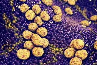
Even more to the point, a herpes virus called cytomegalovirus-- CMV--has long had a suspicious association with accelerated atherosclerosis in heart transplant recipients. And more recent studies have hinted at a link between CMV infection and atherosclerosis in the elderly. So when Epstein, Speir, and their colleagues looked for a culprit, they focused first on cytomegalovirus. Roughly three out of four American senior citizens harbor the virus, usually with no known ill effects. Only in patients with compromised immune systems--such as those with aids, for example, or transplant recipients, who are given drugs to suppress their immune system’s natural rejection of foreign tissue--does the virus pose a proven danger.

Epstein’s team inspected the samples of arterial wall cells collected during the hunt for p53. Was cytomegalovirus lurking there, too? Indeed it was. In fact, 85 percent of the tissue samples from arteries in which there was excessive p53 also had CMV, whereas just 27 percent of samples taken from arteries with normal levels of p53 had CMV in them. That was the first ‘aha,’ recalls Epstein. Finding CMV at the scene of the crime when excess p53 was around implied--but did not prove--that the virus was not just an innocent bystander.

Epstein and his group began to flesh out the following theory. Say the patients had been infected by the virus early in life; it would simply slip silently into their cells and lie low. For years, the virus would get passed from one cell to its progeny. The virus would never be eliminated because it hides in the cell, where the immune system can’t get to it. The cell doesn’t mind, though says Epstein, because nothing bad happens. And the virus, he adds, doesn’t mind because it will still be around 40 years later, when perhaps circumstances will change and it can start to reproduce itself again.

What circumstances allow the virus to reproduce? One known trigger is a heart transplant. Immunosuppressive drugs release CMV from immune system control, and the viral spread is suspected of increasing the risk of cardiovascular complications and rejection. Another possible trigger is angioplasty itself. Expanding the balloon is known to injure the blood vessel, prompting release of factors that promote healing and growth. Epstein suspects that CMV has cunningly adapted to use these chemical signals of injury to turn on viral replication. Studies have shown that the virus, once switched on, begins making a protein called ie84, which binds to p53 and prevents it from working. The result seems to be that infected cells live longer than they should, and their accumulation reclogs the artery.

The data Epstein’s team has collected support this hypothesis. In a study of 75 angioplasty patients last year, Epstein and his colleague Yi- Fu Zhou showed that the CMV-positive patients were much more likely to suffer restenosis. The numbers were very suggestive--reblockage occurred in 43 percent of the 49 patients with CMV. But only 2 of 26 patients who were not infected--less than 10 percent--had suffered reblockage. If you do the statistics, Epstein says, you find that the risk for developing restenosis is nearly 10 times greater in CMV-infected patients.

Still, a strong correlation does not prove causality. Epstein, however, is closing in on more definitive evidence, at least in rats. In a recent study, he and Zhou injured the blood vessels of rats using a procedure similar to angioplasty. The rats were healthy in every way, except that half had been infected with CMV. As the arteries healed, the infected rats developed an excessive accumulation of artery-wall cells that resembled restenosis. The uninfected rats did not. There we have causality, says Epstein. It’s the missing link.

Proving the same in humans will have to wait. The drug ganciclovir is often used to eliminate the threat of CMV infection in heart transplant recipients, but its potential side effects are severe enough that it cannot be used in cases of restenosis, in which the harmful role of CMV infection is not yet proven. At least five drug companies, however, are rushing to develop other, less toxic anti-CMV drugs for patients with AIDS and other diseases marked by weakened immune systems. When one becomes available, there could be clinical trials testing CMV’s role in restenosis.

Epstein feels the evidence, so far, is compelling. As good a case can be made for CMV as was made 15 years ago for cholesterol and its role in atherosclerosis, he says. If CMV becomes accepted as a bona fide risk factor, patients undergoing angioplasty would be tested for CMV and warned that the chances of success are lower if they are infected. More patients might be pushed toward bypass surgery, the primary alternative. Better yet, if researchers come up with a vaccine against CMV, the number of patients developing restenosis might dwindle. But I’m afraid even that is probably too simple, Epstein sighs. I don’t think any single intervention is going to do it. I’ve been in this business too long to think things are going to be so easy.

1 free article left
Want More? Get unlimited access for as low as $1.99/month

Already a subscriber?

Register or Log In

1 free articleSubscribe
Discover Magazine Logo
Want more?

Keep reading for as low as $1.99!

Subscribe

Already a subscriber?

Register or Log In

More From Discover
Stay Curious
Join
Our List

Sign up for our weekly science updates.

 
Subscribe
To The Magazine

Save up to 40% off the cover price when you subscribe to Discover magazine.

Copyright © 2024 LabX Media Group